Technological Innovations
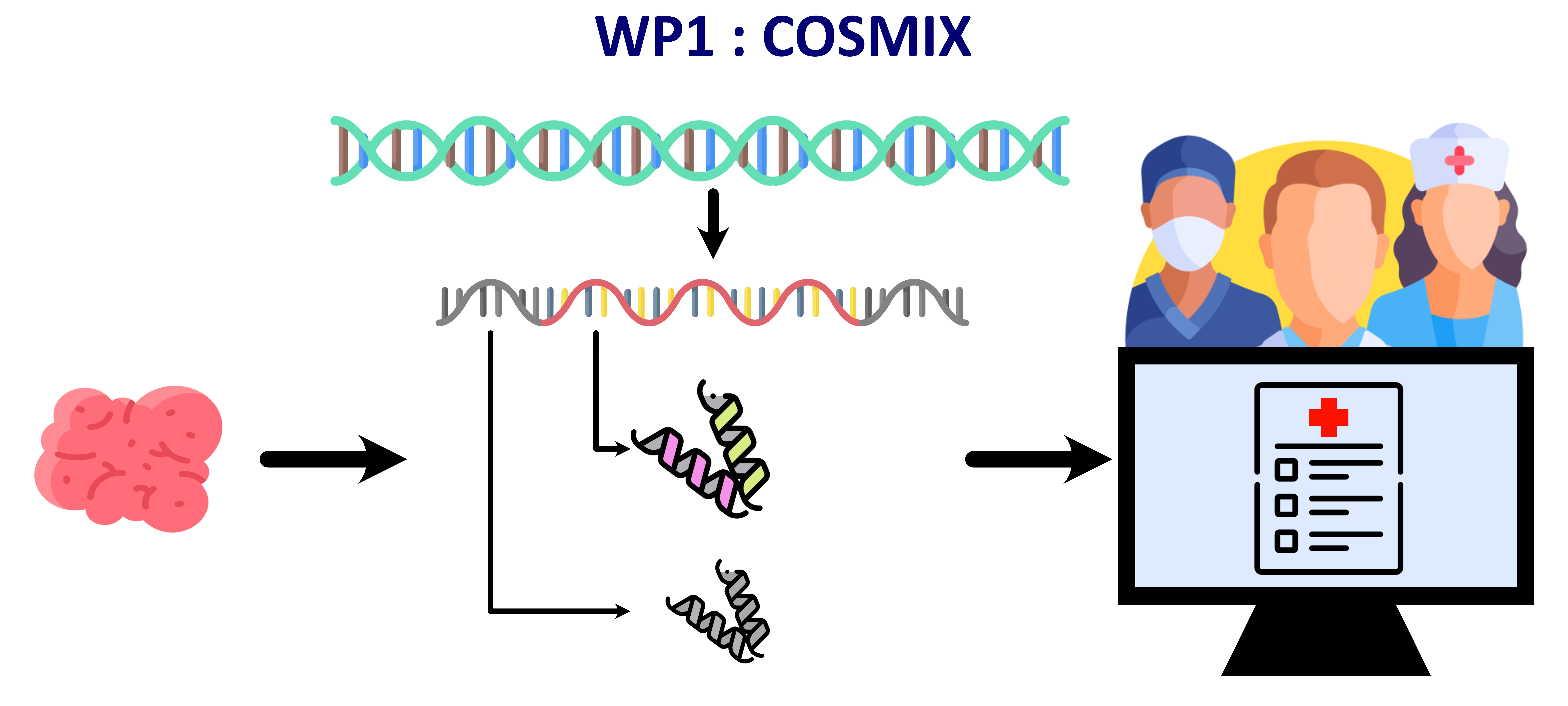
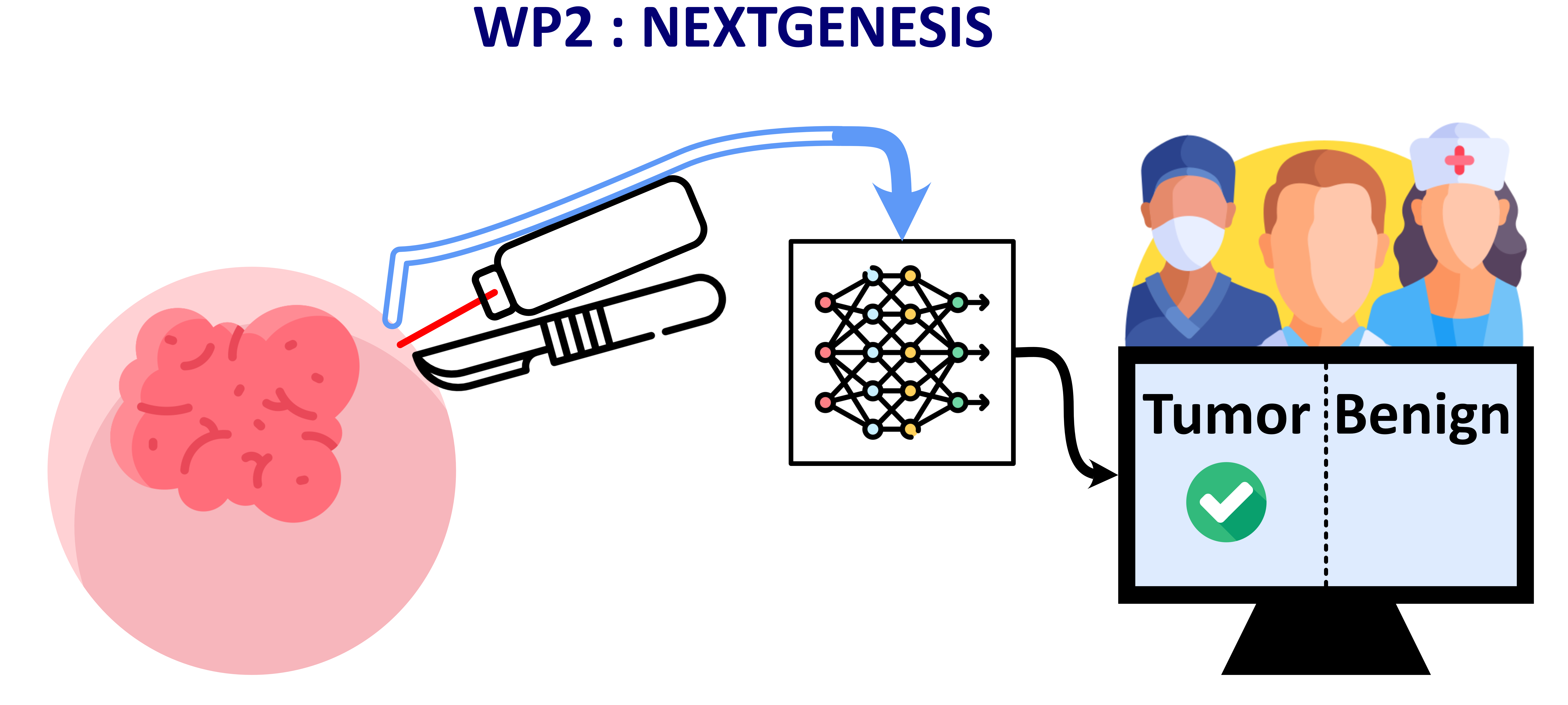
The technological innovation axis is organized in two work packages so-called Early diagnosis and Therapy. The Early diagnosis WP is based on developments of new tools to characterize spatially but also at the single cell and subcellular levels by OMICs the tissue microenvironment from pathological diseases. New captors based on mass spectrometry such SNOOPI for VOCs or BAT-MASS to track these biomarkers in fluids or on patches for diagnosis are currently in clinical trials ‘evaluation. The therapeutic WP is devoted to the next generation surgery through SpiderMass technology including immune mass-based score, in vivo molecular imaging (Deadpool)and photodirect therapy (IrosMass). Several clinical trials using SpiderMass technology are under investigation such as sarcoma, Ovarian Cancer and Breast Cancer (SITH, Maverick, SETH).
Technological innovations

Early Diagnosis

